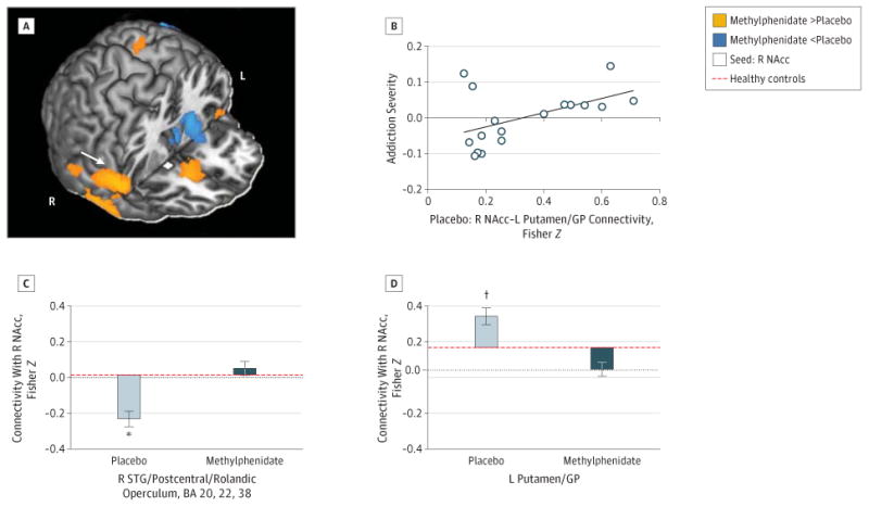
Figure 4. Changes in Nucleus Accumbens (NAcc) Connectivity With Methylphenidate and Relationship to Addiction Severity.

Increased right (R) NAcc (A) (seed shown in white) with R superior temporal gyrus (STG) extending to the postcentral gyrus and rolandic operculum connectivity (C) and decreased R NAcc with left (L) putamen/globus pallidus (GP) connectivity (D) following a single dose of oral methylphenidate hydrochloride, 20 mg. Color map (A) shows increased (orange) or decreased (cyan) connectivity strength with methylphenidate vs placebo in a T-score window from ±3.0 to ±7.0. Bar plots (C and D) show the Fisher Z values for placebo peak effects (light gray) and methylphenidate peak effects (dark gray) plotted from values of healthy control participants scanned during placebo conditions. Error bars represent SEM. The connectivity strength between the R NAcc and L putamen/GP during placebo (C) was uniquely positively correlated with the severity of cocaine addiction composite scores (B). *P < .005. †P < .05.
