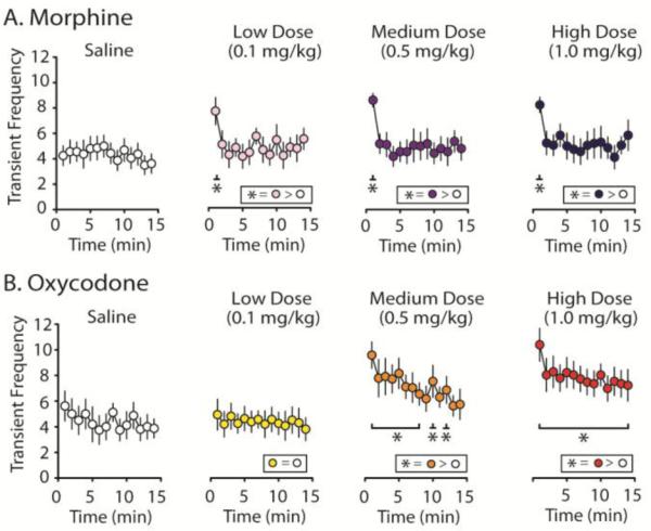
Figure 4.
Dopamine transient frequency following administration of morphine or oxycodone. A. Histology for both morphine subjects (n = 8) recorded from the NAc core (n = 4) or shell (n = 4) and oxycodone subjects (n = 8) - NAc core (n = 4) or shell (n = 4) - were recorded from similar locations within the NAc. Phasic DA transmission data did not differ significantly between the core and shell so data were collapsed. B. The average number of transients per min across the 15 min recording interval are reported following saline controls and cumulatively higher doses of morphine or oxycodone (Low = 0.1 mg/kg, Medium = 0.5 mg/kg, High = 1.0 mg/kg; i.v.). C and D. Transient events were binned every min for the duration of the 15 min recording intervals following each drug dose and compared to the average post-saline frequency. C. Transient data per min following morphine infusions. While morphine failed to affect transient frequency across the entire 15 min recording period, all doses of morphine elicited a significant increase in transient frequency during the first minute following drug infusion compared to saline controls. D. Oxycodone-evoked increases in DA transient frequency were highly dose dependent. While the low dose infusion of oxycodone did not increase transient frequency, the medium dose increased DA transients for most time points and infusion of the high dose of oxycodone elevated transient frequency for the entire duration of the recording period. Error bars indicate SEM. * indicates statistically significant increases (p < 0.05).

