
Fig. 2. Loss of Sept4/ARTS function accelerates wound healing and improves skin regeneration.
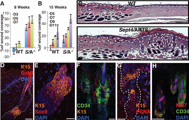
(A and B) Reepithelialization dynamics of skins at different times PWI. Excision wounds (1 cm2) were inflicted on dorsal skin of 8-week-old [(A), n = 12] or 15-week-old mice [(B), n = 8]. Percentage of wound coverage was calculated versus original wound size. D denotes day PWI. (C) Hematoxylin and eosin staining of full-thickness excision wounds of 8-week-old mice, 18 days PWI. Hair follicle formation within the wound bed is observed in Sept4/ARTS−/− mice. (D to F) HFSC niches within regenerated hair follicles are positive for CD34, K15, and Sox9. (D) Regenerated hair follicle displays a HFSC niche positive for K15 and Sox9. (E) Zoom-in of the HFSC niche in (D). (F) Regenerated hair follicle niche is positive for CD34. (G and H) Immunofluorescence for PCNA (G) and Ki67 (H), indicating proliferative activity within the regenerated hair follicle niche of Sept4/ARTS−/− mice. Scale bars, 500 μm (C), 50 μm (D), 20 μm [(E), (F), and (G)], 10 μm (H).