
Fig. 3. Sept4/ARTS−/− HFSCs are responsible for enhanced tissue re-generation.
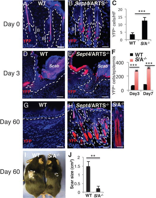
(A to H) Reporter expression was induced from P20 to P25 [(A) to (F)] or from P45 to P50 [(G) and (H)], and wounding was executed at P26 or P56, respectively (see fig. S7). Skins were analyzed at t = 0, 3, 7, 18, and 60 days PWI. WT [(A), (D), (G)] and Sept4/ARTS−/− [(B), (E), (H)] lineage tracings are shown at t = 0, 3, and 60 days. (H), right panel: Close-up of hair follicle at 60 days PWI. Dashed line indicates dermis-epidermis border; B denotes bulge. Scale bars, 50 μm [(A), (B), (G)], 100 μm [(D), (E), (H)]. (C) Quantifications of YFP+ cells in hair follicles of WT and Sept4/ARTS−/− mice (t = 0). (F) Quantification of YFP+ cells in hair follicles and epidermis of WT and Sept4/ARTS−/− mice (t = 3 and 7 days). (I and J) Scar size of WT and Sept4/ARTS−/− mice at 60 days PWI (2 cm2) (n = 3); photograph (I) and quantification (J) are shown. Dashed line indicates scar border. **P < 0.002, ***P < 0.001.