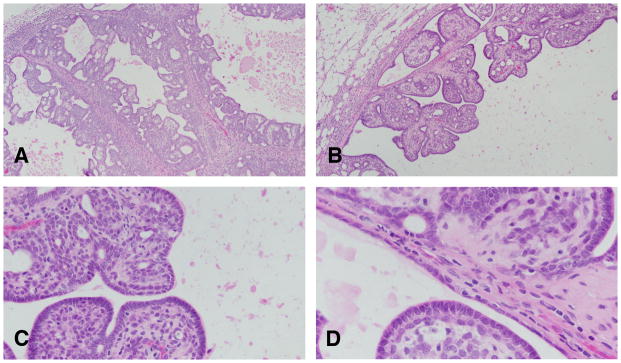
Figure 9.

A: Invasive papillary carcinomas type 1 contain delicate fibrovascular cores, often heavily infiltrated by lymphocytes and mast cells. Stained with H&E, ×4. B: The fibrovascular cores are considerably thinner than those seen in intraductal papillomas. Stained with H&E, ×10. C and D: On top of the fibrovascular core grows the epithelium, which are composed of 1–2 layers of epithelial cells, which in turn emit short epithelial papillae devoid of fibrovascular cores. Stained with H&E, ×40.
