Figure 1.

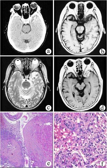
Solid meningioangiomatosis. (a) CT scan showed an irregular mixed high-density mass in the left middle cranial fossa. (b) On T1WI, the lesion demonstrated low and equal signal intensity. (c) On T2WI, the lesion showed high signal intensity with a multiple flow void effect. (d) On post-contrast MRI, the lesion showed significant and homogeneous enhancement. (e, f) Microphotography of specimens showed extensive fibroblastic proliferation and an increased number of vessels surrounded by meningothelial cells.
