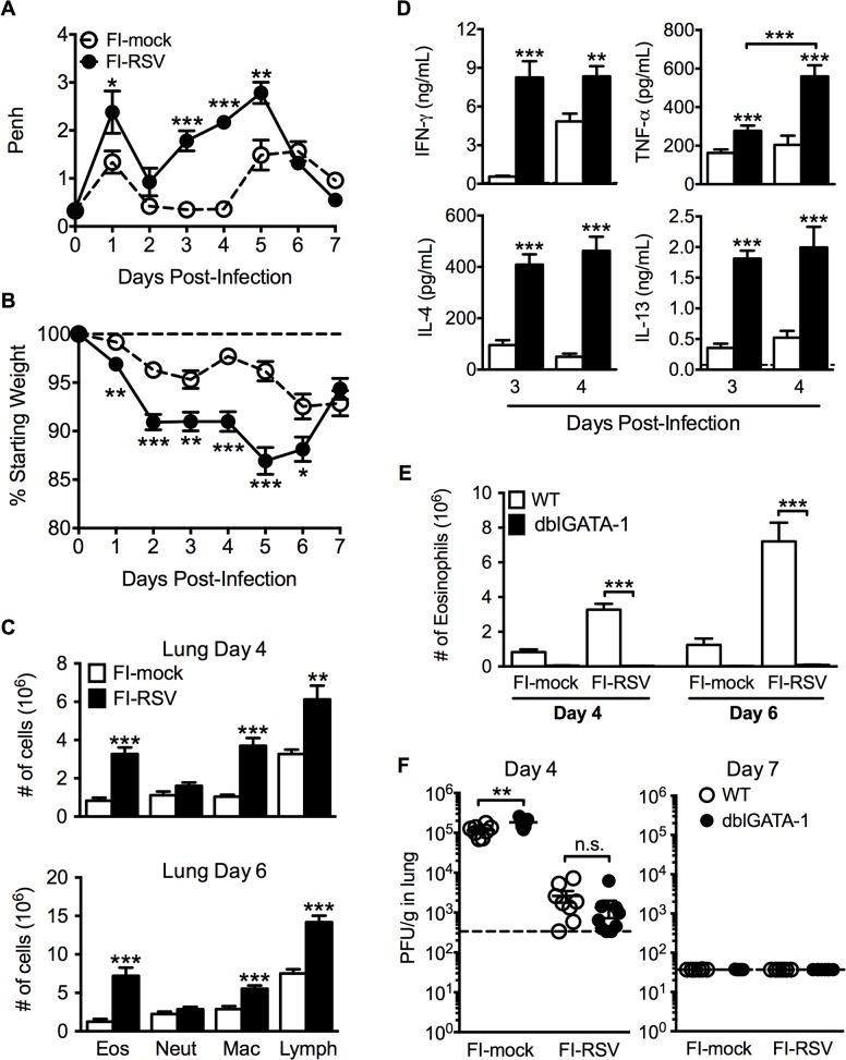
Fig 1. Development of VED following RSV challenge of FI-RSV-immunized mice.
(A) FI-mock- and FI-RSV-vaccinated mice were monitored daily for airway obstruction using a whole body plethysmograph. (B) Weight loss was assessed for immunized mice for 7 days following RSV challenge. (C) Total number of eosinophils (CD11cintSiglec F+), neutrophils (Ly6c+Ly6ghi), macrophages (CD11c+F4/80+), and T cell lymphocytes (CD90.2+) were quantified from the lungs of vaccinated mice via flow cytometry on day 4 and 6 post-RSV challenge. (D) Cytokine protein amounts in whole lung homogenates from immunized mice were determined on day 3 following RSV infection. Dotted lines indicate the limit of detection. (E) Total number of eosinophils from the lung parenchyma of FI-mock- and FI-RSV-immunized WT and dblGATA-1 mice were quantified on days 4 and 6 following RSV challenge. (F) Plaque assay on lungs from immunized WT and dblGATA-1 mice was performed on days 4 and 7 following RSV challenge. Data are represented as mean ± SEM from two independent experiments (n = 12 mice total for A, B, n = 8 for C-F). Groups were compared using Student's t-test for two groups or using one-way ANOVA for comparison of more than two groups, * p<0.05, ** p<0.01, *** p<0.001.

