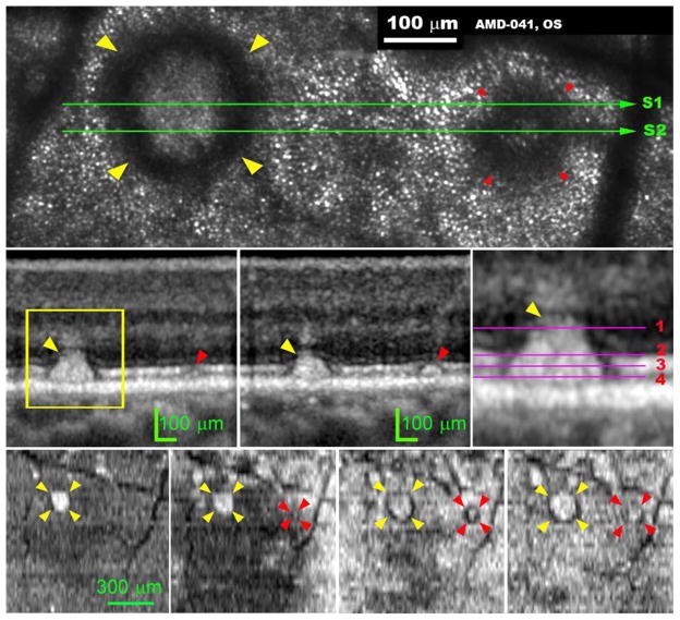
Figure 5.
Subretinal drusenoid deposit shape and location characterized by 3D and high-resolution imaging deposits imaged by adaptive optics scanning laser ophthalmoscopy (AOSLO) and spectral domain optical coherence tomography (SD-OCT). The subject (AMD-041) is a 73-year-old man with non-neovascular age-related macular degeneration (Age-Related Eye Disease Study grade 6, best-corrected visual acuity 20/25). (Top row) AOSLO image, cone photoreceptors are represented by evenly spaced hyper-reflective spots outside the annuli of hyper-reflectivity. The dark vertical bands are the shadows of overlying retinal blood vessels. The yellow and red arrowheads indicate a prominent stage 3 and stage 2 subretinal drusenoid deposits in superior perifovea, respectively, followed through the remaining panels. (Middle row, left and middle) OCT scans through these subretinal drusenoid deposits, as marked by the green line S1 and S2 in the top row panel. The stage 3 subretinal drusenoid deposit lifts the external limiting membrane (ELM) upward and is associated with a crowning tuft of hyper-reflectivity within the inner outer nuclear layer (ONL), representing perturbed Henle fibers.58, 65, 66 The stage 2 subretinal drusenoid deposit indents but does not interrupt the photoreceptor ellipsoidal zone (EZ). (Middle row, right) Magenta lines mark the planes where en-face OCT images, shown in the bottom row panels from left to right, were extracted from the 3D volume OCT scan. AOSLO image was an integration of the light reflected from depth 1 through 4. (Bottom row, far left) The top of a stage 3 subretinal drusenoid deposits has reached the hypo-reflective band internal to ELM. The stage 3 subretinal drusenoid deposit manifests as a highly reflective area on the en face OCT image. (Bottom row, left) at the level near the EZ, the stage 3 subretinal drusenoid deposit becomes larger than at its apex. The stage 2 subretinal drusenoid deposit is barely eligible at this level. (Bottom row, right) at the middle of the EZ, the stage 3 subretinal drusenoid deposit is larger still, and a dark annulus appears; so does the stage 2 subretinal drusenoid deposit. (Bottom row, far right) Near the retinal pigment epithelium (RPE), the hyper-reflective center and hyporeflective annulus of the stage 3 subretinal drusenoid deposit are at their largest. However, the stage 2 subretinal drusenoid deposit is indistinct at this level. The stage 3 subretinal drusenoid deposit shows a wide hyporeflective ring with indistinct borders. The ‘gap’ in the EZ on either side of subretinal drusenoid deposits in the OCT B-scan corresponds to the hyporeflective annulus on the en-face OCT and the AOSLO image.

