
Figure 5.
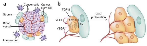
Signaling pathways in skin cancers. (a) The squamous cell carcinoma–cancer stem cell niche. CSCs are often found at the tumor-stroma interface, together with an elaborate vasculature, immune cells and aberrant fibroblasts. (b) Upon activation of αβ1 integrins by extracellular matrix ligands such as fibronectin (FN), focal adhesion kinase (FAK) and its associate tyrosine kinase Src become hyperactivated and promote proliferation of CSCs. By contrast, TGF-β signaling counteracts integrin activity and enhances CSC quiescence. In addition, CSCs secrete VEGF, which acts in an autocrine fashion to enhance CSC proliferation and in a paracrine fashion to promote formation of new blood vessels.