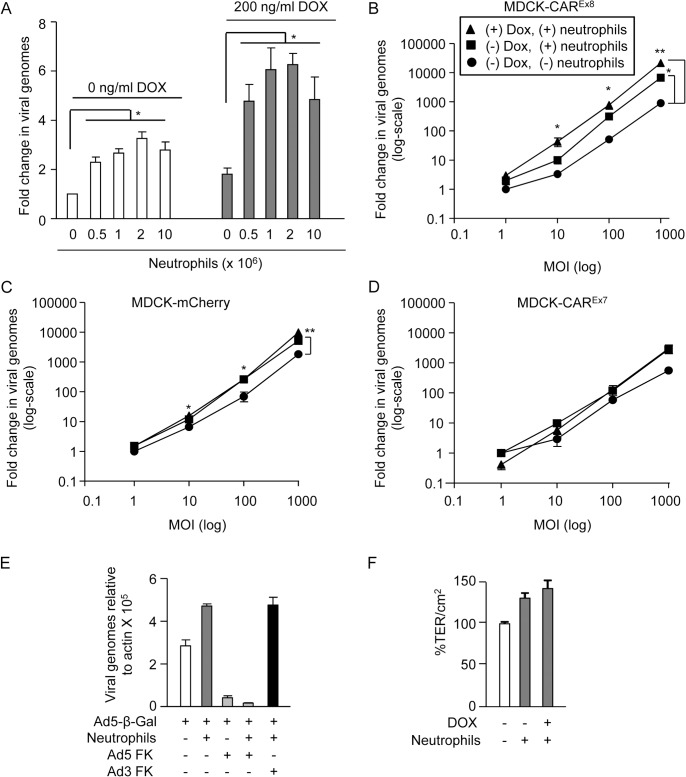
Fig 5. Neutrophils adhered to the apical surface of polarized-MDCK cells augment AdV entry without decreasing the TER.
A) MDCK-CAREx8 cells were either mock- or DOX-induced. A neutrophil adhesion assay was performed with increasing numbers of neutrophils, as indicated. Immediately post-neutrophil adhesion, MDCK-CAREx8 epithelia were infected with AdV5-β-gal for 1 h from the apical surface. 24 h later, viral entry was determined by qPCR analysis. Fold change in viral genomes, relative to AdV5-βGal entry in the absence of DOX and neutrophils, is shown. AdV entry from the apical surface was quantitated by qPCR analysis of polarized B) MDCK-CAREx8 C) MDCK-mCherry and D) MDCK-CAREx7 cells that were uninduced (circles), uninduced with adhered neutrophils (squares), or induced with DOX for 24 h prior to neutrophil adhesion (triangles). E) AdV5-β-gal entry from the apical surface of MDCK-CAREx8 epithelia in the presence or absence of neutrophils and AdV5 FK or AdV3 FK. F) TER of mock- or Dox-induced MDCK-CAREx8 epithelia was measured in the presence or absence of neutrophils. Error bars represent standard error of the mean (SEM) from three independent experiments. No significant difference was detected by one-way ANOVA. Error bars represent the SEM from three independent experiments; *p < 0.05 or **p < 0.001 by one-way ANOVA and Bonferroni post hoc test.

