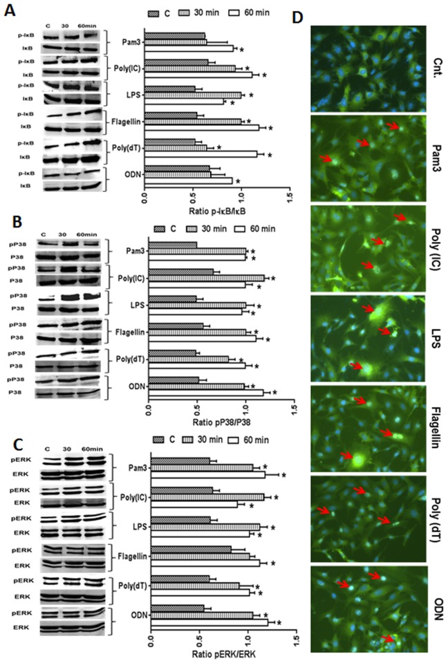
Fig 3. Activation of TLR-downstream (NF-kB and MAPK) signaling following TLR agonist stimulation.
In order to assess the activation of IkB, p38, and ERK signaling following stimulation with TLR agonists for 30 and 60 min., 661W cells were lysed in RIPA buffer and analyzed by Western blot analysis using antibodies against phospho-IkB (p-IkB), p-p38, and p-ERK. Antibodies against ERK, p38, and IkB were used to detect total protein levels (A-C). Band intensity was quantified using Image J and presented as the relative band intensity of the phosphorylated form vs. the total form for the respective proteins (A-C). To detect the nuclear translocation of NF-kB (p65), 661W cells were challenged for 60 min. with various TLR ligands. Cells were then fixed, permeablized, and immune-stained with antibodies against p65, a subunit of NF-kB (D). Statistical analysis was performed using one-way ANOVA (*, p<0.05), for comparisons of control versus 30 and 60 min. stimulation.

