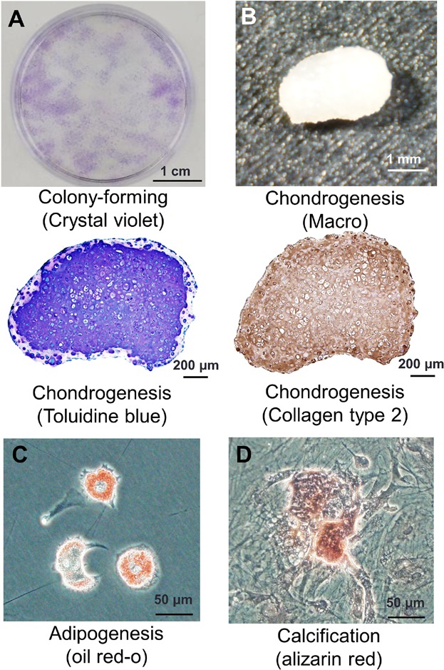
Figure 3.

Colony-forming ability and multipotentiality of the cells derived from IFPs. (A) Representative cell colonies stained with crystal violet. (B) Cartilage pellet and its histological sections stained with toluidine blue and type 2 collagen. (C) Adipocytes stained with Oil Red-O. (D) Calcification stained with alizarin red.
