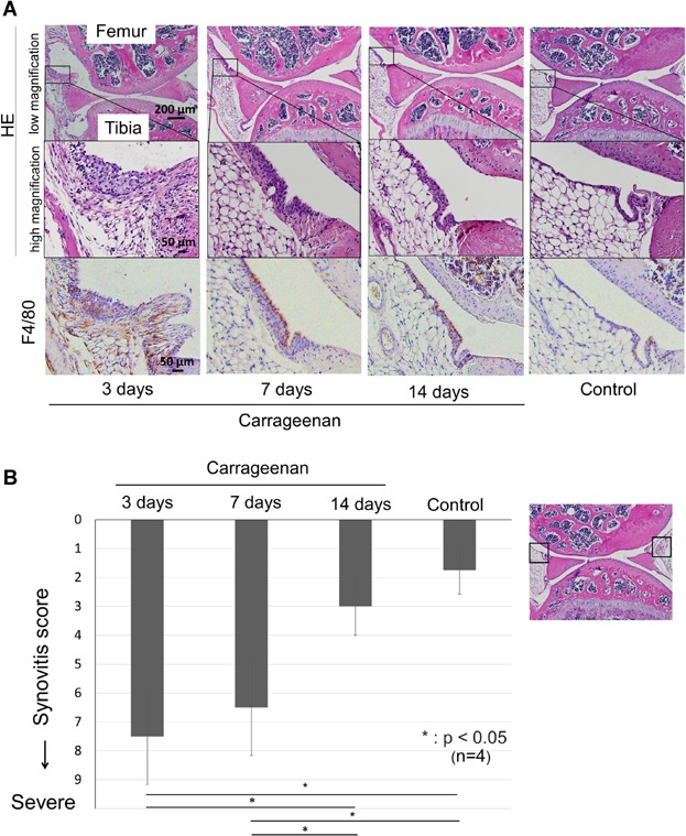
Figure 5.

Histological analyses for synovitis induced by carrageenan. (A) Sagittal sections of mice knee joint, stained with H&E and F4/80 antibody for macrophage, 3, 7, and 14 days after injection of carrageenan and as a control. (B) Synovitis score. Severity of synovitis evaluated by scoring system. Average values with standard derivation are shown. For the synovitis score described by Krenn et al.,19 anterior and posterior synovium was evaluated as denoted in the right panel (*p < 0.05 by Tukey–Kramer test).
