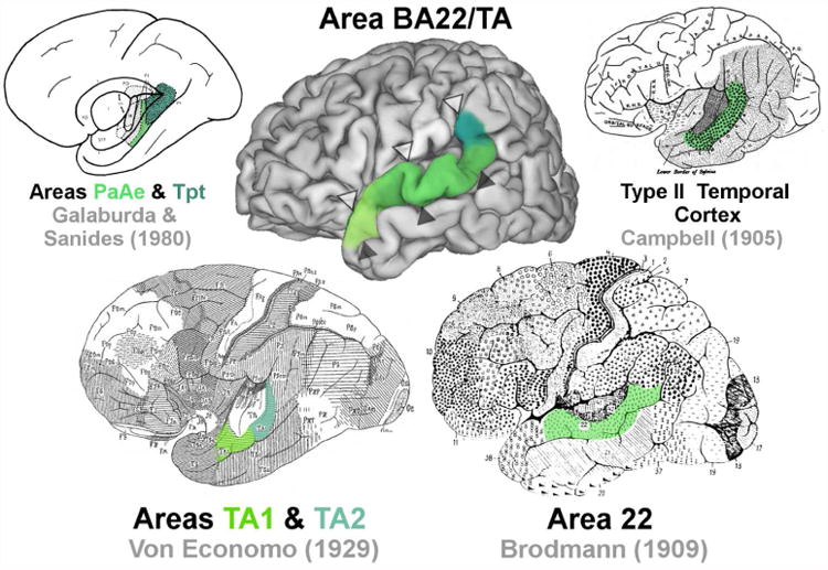
Figure 1.

A map of the approximate location of BA22/TA (middle) compared with classical maps of the region. Open arrowheads (Δ) indicate the position of the lateral sulcus. Closed arrowheads indicate the position of the superior temporal sulcus (STS) (▲).
