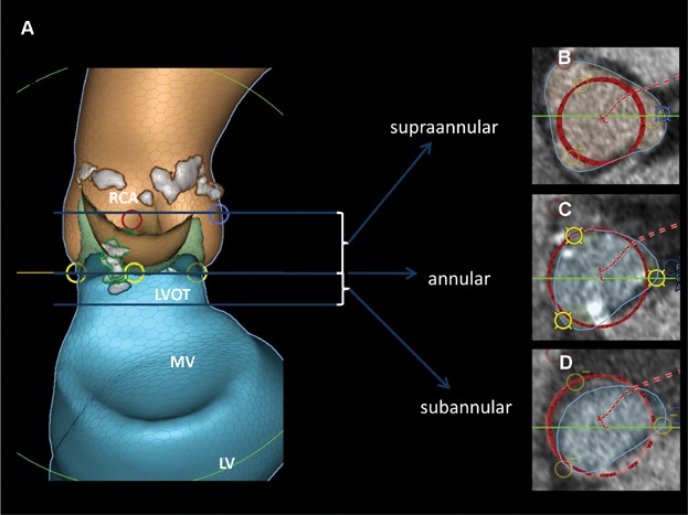
Figure 2.

Preimplant analysis. (A) LCA and blue circle, origin of the left coronary artery; RCA and red circle, origin of the right coronary artery; yellow circle, nadir of the cups; LVOT, left ventricular outflow tract; MV, mitral valve region; LV, left ventricle; grey area, calcifications. (B–D) Blue circle, cross sectional area of region of interest; red circle, valve dummy in the region of interest.
