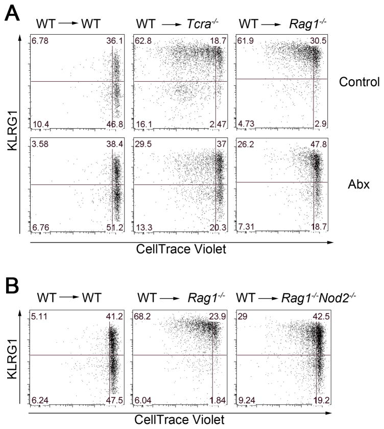
Figure 4. Commensal bacteria are partially responsible for homeostatic proliferation of NK cells and induction of KLRG1 expression.
(A) Enriched WT (CD45.1+) NK cells were labeled with CellTrace Violet and 5 × 10⁵ cells were transferred via intravenous injection into WT mice (CD45.2+), Tcra−/− mice (CD45.2+), or Rag1−/− mice (CD45.2+) and given either control or Abx-treated water for 14 days prior to transfer. (B) CellTrace-Violet-labeled WT NK cells were transferred into WT mice (CD45.2+), Rag1−/− mice (CD45.2+), or Rag1−/−Nod2−/− mice (CD45.2+). KLRG1 expression and dilution of CellTrace-Violet in splenic CD45.1+ NK cells were analyzed 7 days after transfer. Data are representative of three independent experiments with 3 mice in each experiment. See also Figure S4.

