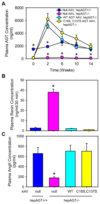
Figure 2. Deletion of Cys18-Cys137 disulfide bond of AGT did not affect plasma concentrations of AGT, renin, or AngII.
(A) Plasma total AGT concentrations were measured using an ELISA kit at baseline, and 2, 6, 10, and 14 weeks after AAV injection (N = 4 – 5/group). Statistical analyses were two way repeated measures ANOVA. * denotes P < 0.001 versus the other 3 groups at each time point. (B) Plasma renin concentrations were measured at termination by radioimmunoassay (N = 7 – 10/group). * denotes P < 0.001 versus the other 3 groups by one way ANOVA and post-hoc comparison of Tukey-Kramer adjustment. (C) Plasma AngII concentrations were measured at termination by radioimmunoassay (N = 3 – 5/group). * denotes P = 0.002 versus the other 3 groups by one way ANOVA with Holm-Sidak method. WT represents wild-type AGT AAV, and C18S,C137S represents AGT in AAV vector with Cys to Ser mutation at 18 and 137 residues.

