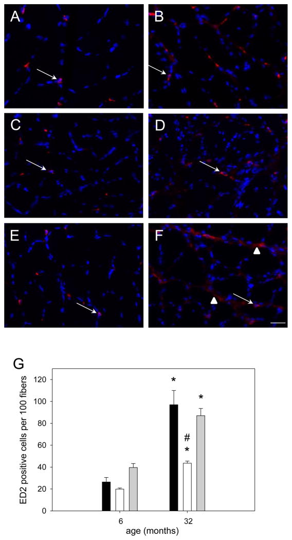
Figure 13. Macrophage (ED2) abundance in dysregulated in soleus muscle of aged rats.
Representative cross sections immunoreacted for ED2 (CD163) of soleus muscle from 6 (A,C,E) or 32 (B,D,F) month old ambulatory (A,B), hind limb suspended (C,D), or reloaded (E,F) rats. CD163 is stained in red and nuclei are stained in blue (DAPI); arrows indicate regular staining and arrow heads point to diffuse CD163 positive staining. Bar in F represents 25μm for all pictures. Quantification of ED2 positive staining (G) in soleus muscle from ambulatory (amb, black bars), hind limb suspended (HS, white bars) and reloaded (HSRE, grey bars) rats at 6 and 32 months is shown (n=8–10). Values are means ± SE; * indicates a significant difference from young and # indicates a significant difference from ambulatory and HSRE in old; p<0.05.

