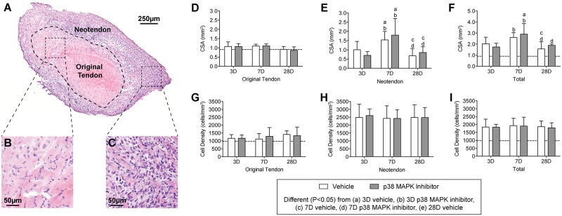
Fig 3. Representative cross-sections of plantaris tendons subjected to synergist ablation, stained with hematoxylin and eosin.

(A) Low magnification view of the tendon, and high magnification views of (B) the original tendon and (C) the neotendon are shown. (D-I) Quantitative analysis of tendon sections: Cross-sectional area (CSA, in mm2) of (D) the original tendon, (E) the neotendon, and (F) the total tendon. Cell density (cells/mm2) of (G) the original tendon, (H) the neotendon, and (I) the total tendon. Values are mean±SD. N, 4 to 8 tendons for each group. Differences between groups were tested using a two-way ANOVA (α = 0.05) followed by Newman-Keuls post hoc sorting: a, different (P<0.05) from 3D vehicle; b, different (P<0.05) from 3D p38 MAPK inhibitor; c, different (P<0.05) from 7D vehicle; d, different (P<0.05) from 7D p38 MAPK inhibitor; e, different (P<0.05) from 28D vehicle. For reference, horizontal dashed line indicates wet mass of plantaris tendons that were not subjected to synergist ablation.
