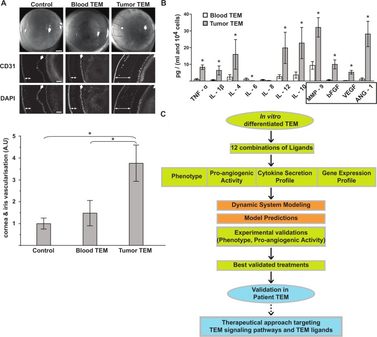
Fig 1. Phenotypical signature of pro-angiogenic TEM.
(A) In vivo corneal vascularization assay to assess the pro-angiogenic activity of TEM isolated from peripheral blood and tumor of breast cancer patients. Bright field pictures of the eyes and fluorescent microscopy images of sagittal sections of the eyes stained with CD31 (stains specifically blood vessel endothelial cells) and Dapi (stains cell nucleus) are shown. Double-head and single arrows depict cornea and iris respectively. Corneas of control eyes were injected with buffer alone and show similar vascularization to uninjected eyes. Note the presence of blood vessels in the cornea injected with tumor TEM (double-head arrow) and its absence in the corneas injected with buffer (control) or blood TEM. Shown are representative data of 10 experiments. Bars in bright field and fluorescent images are 500 and 100 μm respectively. Bar graph represents a quantification of the vascular network of the cornea and the iris. (B) Secretion profile of cytokines and angiogenic factors in TEM isolated from patient blood and tumor. Angiogenic factors are boxed. Shown are cumulated data of 5 experiments, significant variations (P < 0.05) are indicated with an asterisk. (C) Workflow diagram of the strategy combining experimental and computational approaches to discover anti-angiogenic therapies. Green: experiments using ivdTEM. Blue, experiments using patient TEM; red: computational approach.

