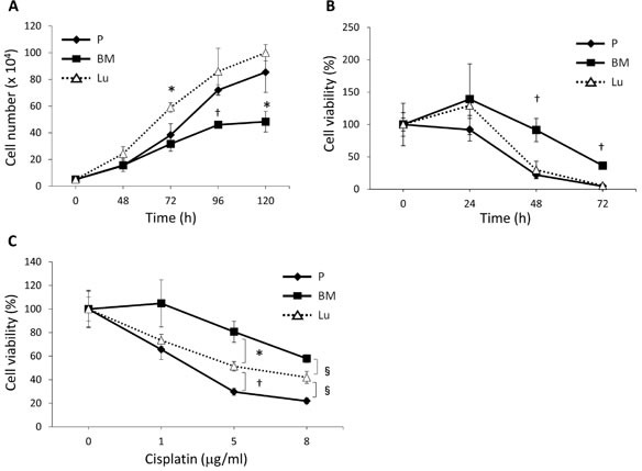
Figure 2. Cell proliferation and cisplatin sensitivity of BM-derived and lung-derived DTCs.

(A) Proliferation rate of P-HEp3 (P), Lu-HEp3 (Lu), and BM-HEp3 (BM) cells. The number of cells in each subline was determined at the indicated time points after plating. *P < .05, †P < .01 compared with P-HEp3 cells. (B) Cells were serum-starved and then their survival was evaluated at the indicated time points. †P < .01 compared with P-HEp3 and Lu-HEp3 cells. (C) Cells were treated with cisplatin at the concentrations shown for 48 hours, after which cell numbers were determined. *P < .05, †P < .01,§P < .005. Values are means ± SEM of triplicate samples.
