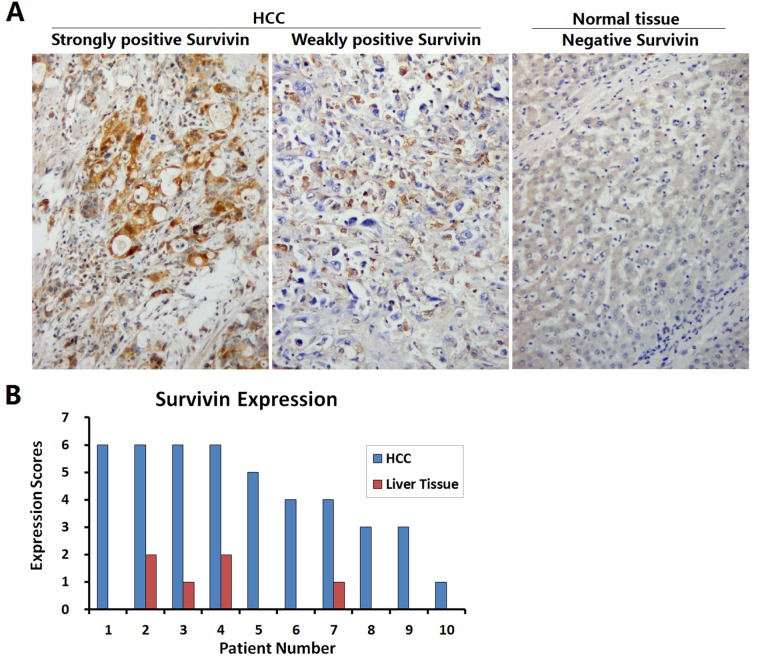
Figure 2. Identification of Survivin expression in HCC specimens.
(A) HCC specimens and paracancerous liver tissues collected from 10 patients with HCC were fixed in 10% formalin for 6 h to prepare the paraffin-embedded sections, and the expression of Survivin was detected by streptavidin-peroxidase (SP) immunohistochemistry; original magnification: ×200. (B) For each slice, the number of Survivin positive cells was counted within 5 medium-power magnification fields of view (20× objective lens) under microscope. The results were determined by scoring the stained cell ratio and the staining intensity, from 0 to 6 scores.

