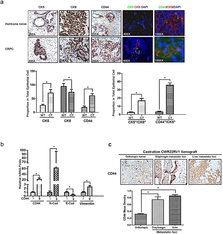
Figure 5. CD44+ stem-like cells are responsible for mesenchymal transition and metastasis.
(a) The increasing expression of CK5 and CD44 in human CRPC samples comparing with hormone naïve PCa was demonstrated in IHC staining (three lanes on the left), immunofluorescence double staining of CK5 and CK8(the fourth lane from left), and immunofluorescence double staining of CD44 and CK8 (the fifth lane from left). (b) CD44+ and CD44− LNCaP cells were separated by MACS, their CD44, E-cadherin, N-cadherin and Vimentin expression were detected by real-time PCR. (c) CWR22rv1 cells were orthotopically implanted into the anterior lobes of nude mice prostate to generate xenograft tumors. CD44 expression in liver and diaphragm metastatic foci were compared to orthotopical xenograft tumors in IHC assay. Quantitation was shown in the right. Significance was defined as p<0.05(*).

