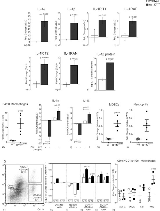
Figure 1. mRNA expression of IL-1RT1 ligands and associated proteins in the distal stomach of 30 week old gp130757FF compared to wildtype mice.

(A)(i) IL-1α, (ii) IL-1β, (iii) IL-1R type 1, (iv) IL-1RAP, (v) IL-1R type 2 and (vi) IL-1RAN. (B) Protein expression of (i) IL-1β in the distal stomach of 30 week old gp130757FF compared to wildtype mice. (C) Proportion of F4/80 macrophages in the stomach of 12 week gp130757FF mice compared to wildtype mice. (D) mRNA expression of (i) IL-1α and (ii) IL-1βin peritoneal macrophages isolated from either wildtype or gp130757FF cultured in the presence or absence of LPS (100ng/μl). (E) Proportion of (i) MDSC and (ii) neutrophils in the stomach of 12 week gp130757FF mice compared to wildtype mice. (F) Presentation of how CD45+ gastric immunocytes were sorted for CD11b and Gr1. (G) mRNA expression of IL-1α and IL-1β of cell sorted populations; (i) unsorted cells, (ii) CD45+ /CD11b−, (iii) CD45+ /CD11b+ /Gr1− and (iv) CD45+ /CD11b+ /Gr1+. (H) mRNA expression of M1 macrophages markers (TNF-α and iNOS) and M2 macrophage markers (Ym1 and Ym2). Bars are means ± SEM, p-values are presented for statistically significant changes (p < 0.05).
