
Figure 4. Pro-angiogenic functions of exosome-differentiated BM-MSC.
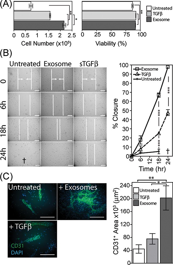
Conditioned media from BM-MSC pre-treated for 4 days as specified, was added to wells containing 1 × 10⁴ primary HUVEC. Following 6 days in culture, HUVEC were harvested and viability and cell counts performed using the ViaCount system on a Guava flow cytometer (Bars, mean ± SD, of triplicates) (A). Confluent monolayers of HUVEC were established, prior to scrapping a single vertical scratch using a 200μl pipette tip. CM from BM-MSC pre-treated as specified, were added, and the distance between two sides of the scratch, highlighted by vertical white lines and arrows, was monitored at specified time points microscopically up to 24 h. The symbol † depicts a loss of HUVEC adhesion at 24 h, hence scratch width could not be measured. (Graph shows Mean ± SD, of duplicate wells per treatment. Data are representative of two such experiments (B). Monolayers of BM-MSC were pre-treated as specified for 4 days, at which point 50% of the culture medium was removed, and replaced by the same volume of EBM2 endothelial cell culture medium lacking growth factors, containing 20,000 HUVEC per well. After further 6 day incubation, the co-cultures were fixed and immune-fluorescently stained for CD31 (green) and DAPI (blue) (Scale, 100μm). Quantification of surface area of the CD31-positive structures was performed (Bars, Mean ± SD of triplicate wells per condition). Representative of three such experiments (C).