
Figure 2. Overexpression of p16INK4A and Ki-67 increases with progressing severity of cervical lesions.
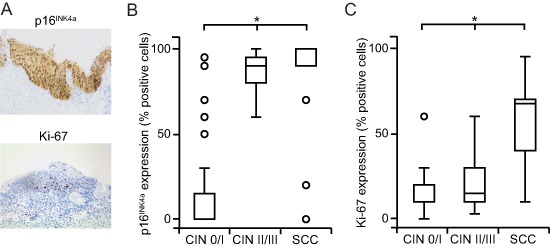
(A) Cells immunohistochemically positive for p16INK4A show a strong nuclear and cytoplasmic expression; cells positive for Ki-67 exhibit an intense nuclear labeling with a limited diffuse background staining (representative bright field images, 20x magnification). (B, C) The extent of staining for p16INK4A (B) and Ki-67 (C) is higher in in SCC and CIN II / III compared to CIN 0 / I cohorts. p16INK4a and Ki-67 expression was recorded as the percentage of positive cells among the total number of epithelial cells in representative areas and stratified by histopathology. Box plots summarize the median, the 25th and 75th percentiles, the whiskers and the outliers (*p < 0.05, after Bonferroni correction).