
Fig.3. Characterization of T cell subsets of OT-I origin within tumors.
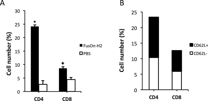
Tumors were explanted from mice used in Fig.2, which had been treated either with PBS (mock) or FusOn-H2 before adoptive transfer of OT-I cells. After the Histopaque density gradient separation of cell suspension was prepared from the harvested tumors, the layer containing lymphocytes and monocytes was collected and stained for CD4 and CD8 (A) or CD4/CD62L and CD8/CD62L (B), respectively. The data in B is from tumor treated with FusOn-H2. Due to the low number of CD4 and CD8 T cells that could be harvested from the tumor treated with PBS, the number of CD4/CD62L and CD8/CD62L subpopulations was insufficient for a reliable quantitative measurement by flow cytometry. The percentage represents the positively stained cells out of the total cells enriched within the layer. +p<0.05, *p<0.01 as compared with PBS.