
Figure 2. Overexpression of FGF8 correlates with lymph node metastasis and poor prognosis in CRC.
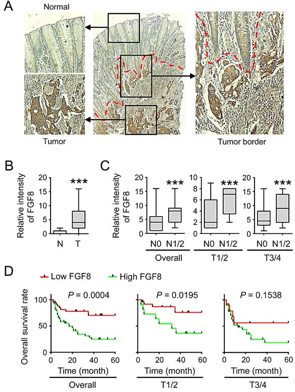
(A) Immunohistochemical staining of FGF8 in tumor and corresponding colorectal mucosa. (B) Immunohistochemical scores for FGF8 in normal colorectal mucosa and CRC tissues. (C) Expression of FGF8 in the primary tumors without (N0) or with (N1/N2) lymph node metastasis was analyzed. Left, overall tumors; middle, stage T1–T2; right, stage T3–T4. (D) Kaplan–Meier survival curves of CRC patients with low (n = 34) and high (n = 38) FGF8 expression. Left, overall tumors; middle, stage T1–T2; right, stage T3–T4. ***, P<0.001.