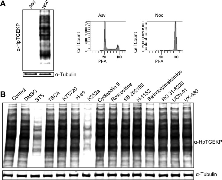
Figure 1. K252a can inhibit linker phosphorylation in mitotic HeLa cells.
(A) Western blot analysis of whole protein extracts from HeLa cells, growing asynchronously (Asy) or arrested with nocodazole (Noc) for 18 hours. The blot was probed with anti-HpTGEKP antibody to visualize the C2H2 ZFP linker phosphor-bands. The blot was then probed with anti-tubulin to show equal loading. A fraction of the cells were tested with FACS analysis for their cell cycle distribution, as a control (right panels) (B) HeLa cells arrested with nocodazole for 18 hours were collected, divided into equal fractions and treated with small-molecule inhibitors, as indicated on the blot, at 1 μM concentration for 10 minutes. Cell lysates were analyzed by Western blotting and probed with anti-HpTGEKP antibody, then with anti-tubulin antibody as loading control.

