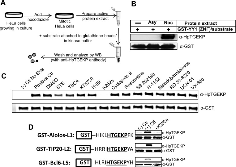
Figure 2. K252a can inhibit the linker kinase activity in mitotic extracts in vitro.
(A) Experimental outline for in vitro kinase assays using active mitotic protein extracts. (B) Western blot analysis of in vitro kinase assay performed as described in (A) using GST-YY1 (ZNF) as substrate coupled to glutathione beads. The blot was probed with anti-HpTGEKP antibody to show phosphorylation by mitotic extracts and anti-GST antibody to show equal substrate loading. (C) Protein extracts from nocodazole-arrested HeLa cells were tested in an in vitro kinase assay as described in (A) and (B) in the absence or presence of the indicated small molecule inhibitors. (D) The mitotic protein extracts were further tested in in vitro kinase assays with three GST-tagged linker sequences from three different proteins (as indicated), coupled to glutathione beads. The assays were performed in the absence or presence of K252a. The Western blots were analyzed by anti-HpTGEKP antibody, then with anti-GST antibody to show equal substrate loading.

