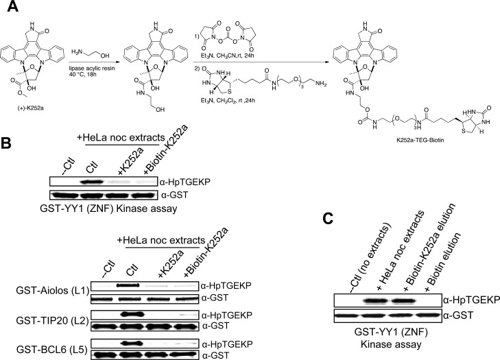
Figure 3. Biotinylated K252a maintains linker kinase inhibitory activity.
(A) Outline of the preparation of biotin-K252a compound. (B) Western Blot analysis of the in vitro kinase assays testing the activity of protein extracts from nocodazole-arrested HeLa cells on GST-tagged linker sequences, coupled to glutathione beads, in the absence or presence of K252a or biotin-K252a. (C) Biotin-K252a (or biotin as negative control) was incubated with protein extracts from nocodazole-arrested HeLa cells, followed by incubation with avidin beads. After collecting the beads by centrifugation and washing, the protein fraction bound to biotin-K252a was eluted with ATP, and tested for linker kinase activity in an in vitro kinase assay with GST-YY1(ZNF) as substrate. The kinase reactions were analyzed by Western blotting with anti-HpTGEKP and anti-GST antibodies. Biotin pull-down served as negative control and the total mitotic protein extracts as positive control.

