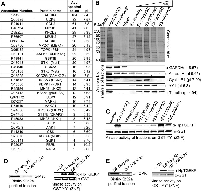
Figure 4. Identification of TOPK as a primary linker kinase candidate.
(A) List of the kinases physically associating with biotin-K252a (but not biotin only) in the avidin pull-down assay, as identified by mass spectrometry analysis. The kinases are listed according to the average spectral counts from the three independent pull-downs. The list of the respective isoelectric point is also included for each kinase. (B) Step-wise anion exchange fractionation of protein extracts from nocodazole-arrested HeLa cells. The proteins were bound to the resin of a spin column and then eluted at the indicated NaCl concentrations. Samples from the input extract, wash, and fractions were run on SDS-PAGE gels and either stained with Commassie blue or Western blotted and probed with the indicated antibodies. (C) Samples were also tested in in vitro kinase assay with GST-YY1(ZNF) coupled to beads, as substrate. The kinase reactions were Western blotted and probed with anti-HpTGEK and anti-GST antibodies (please also refer to Figure S4). (D–E) An elution fraction of a biotin-K252a pull-down was immunodepleted overnight with anti-Mst1/2 antibody or anti-TOPK antibody; the negative control depletion was incubated with a negative rabbit antibody (D and E left panels). The Mst1/2 or TOPK immunodepleted biotin-K252a fractions were tested in in vitro kinase assays with GST-YY1(ZNF) coupled to beads. The kinase reactions were Western blotted and probed with anti-HpTGEKP and anti-GST antibodies (D and E right panels).

