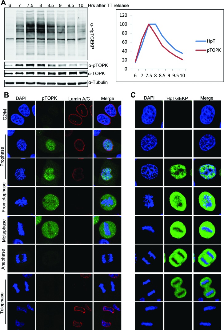
Figure 6. Timing and localization of mitotically active pTOPK in correlation with HpTGEKP.
(A) HeLa cells were synchronized at G1/S by double-thymidine block and then released. Cells were collected for whole cell extract preparation at 6–10 hours after the release (as indicated), as they entered and exited mitosis. Protein extracts were analyzed on a Western blot with anti-HpTGEKP, anti-TOPK, and anti-pTOPK antibodies. The blot was further probed with anti-Tubulin as a loading control. The signals from the anti-HpTGEKP and anti-pTOPK antibodies were quantified and plotted, after setting the highest signal from each antibody to 100. (B-C) HeLa cells grown on coverslips were synchronized with a single thymidine block and then released (right panel). Cells were fixed 7–9 hours after the release, permeabilized, and immunocytostained with anti-pTOPK, anti-Lamin A/C (to visualize the nuclear envelope) (B), and anti-HpTGEKP (C) antibodies. Cells were stained with DAPI to visualize the DNA. Please refer to Figure S6 (for total TOPK immunostaining) and Figure S8 (for pTOPK immunostaining in multiple mitotic cells in the same field).

