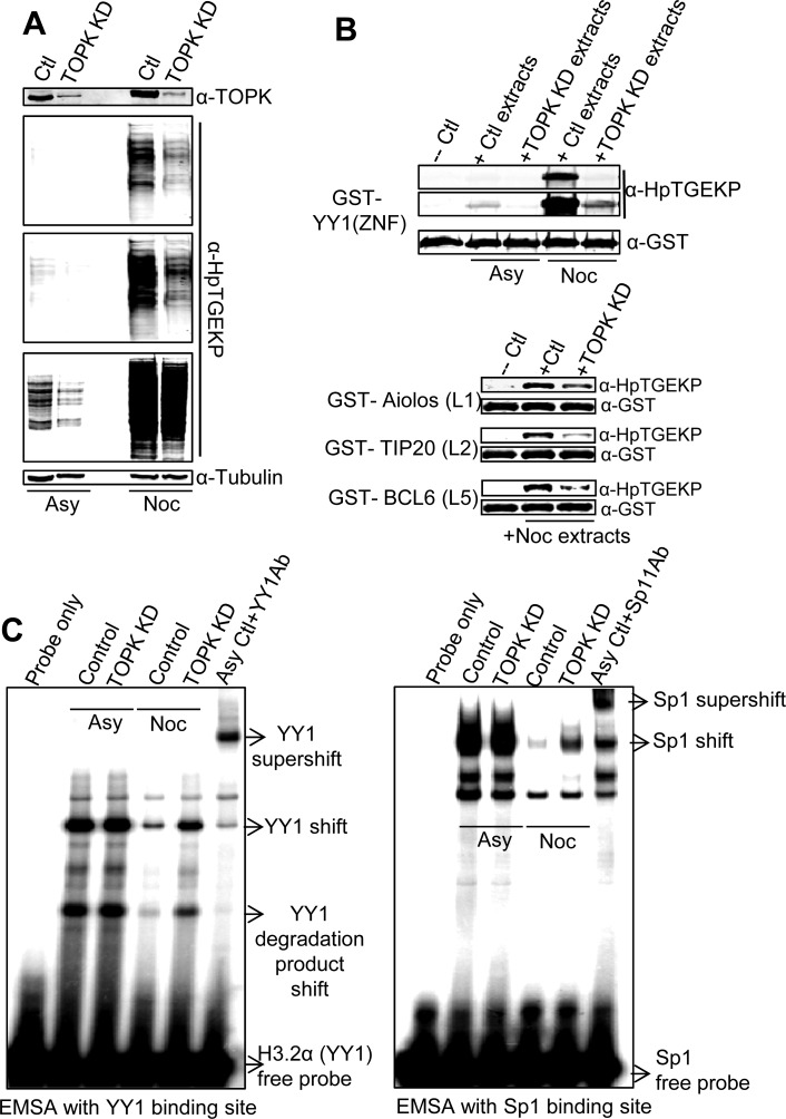
Figure 7. TOPK knockdown results in significant reduction of linker phosphorylation in vivo.
HeLa cells were transfected with TOPK specific siRNA, or scrambled siRNA, and either left growing asynchronously or synchronized with thymidine-nocodazole block. (A) Western blot analysis of WCE of siRNA transfected HeLa cells probed with anti-TOPK and anti-HpTGEKP antibodies. Three different exposures of the HpTGEKP signal are displayed to visualize the differences in HpTGEKP signal between control and TOPK knockdown samples, both in asynchronous and mitotic cells. The blot was further probed with anti-Tubulin as a loading control. (B) The proteins extracts probed in (A) were tested in an in vitro kinase assays with purified GST-tagged: YY1(ZNF), Aiolos (L1), TIP20(L2), or Bcl6(L5) as substrates. The kinase reactions were analyzed by Western blotting with anti-HpTGEKP and anti-GST antibodies. (C) The proteins extracts probed in (A) were tested in in vitro EMSA assays by incubation with radioactively-labeled double-stranded DNA oligonucleotides comprising the YY1 and Sp1 consensus binding sites. The YY1 and Sp1 specific shifts are indicated and confirmed by super-shift analysis with their respective specific antibodies.

