
Figure 4. GANT61 downregulates GLI1, GLI2 and PTCH1 through ROS.
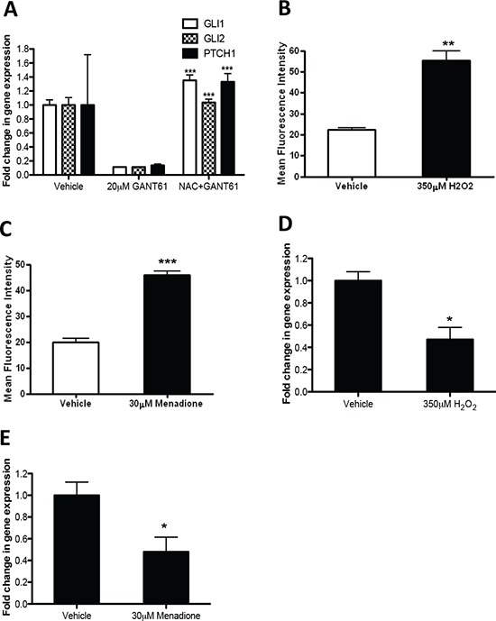
(A) Analyses of Hh pathway genes (GLI1, GLI2 and PTCH1) by qRT-PCR. LO68 cells were pretreated with NAC for 1 h and then with 20 μM GANT61 or vehicle for 48 h. Values represent the mean ± SEM of three independent experiments each performed in duplicate. ***, p < 0.001, compared to GANT61-treated cells. (B) LO68 cells were treated with vehicle or 350 μM H2O2 for 24 h. The cells were stained with CH2DCFDA and the fluorescence measured by flow cytometry. Bar graph represents the increase in mean fluorescence intensity of positive cells measured in each experimental condition. Values are the average of independent measurements (mean ± SEM; n = 3). **, p < 0.01, compared to vehicle-treated cells. (C) LO68 cells were treated with vehicle or 30 μM menadione for 24 h. The cells were stained with CH2DCFDA and fluorescence measured by flow cytometry. Bar graph shows the increase in the mean fluorescence intensity of positive cells measured in each experimental condition. Values are the average of independent measurements (mean ± SEM; n = 3). ***, p < 0.001, compared to vehicle-treated cells. (D) Analysis of GLI1 mRNA expression by qRT-PCR. LO68 cells were treated with vehicle or 350 μM H2O2 for 24 h. Values represent the mean ± SEM of three independent experiments each performed in duplicates. *, p < 0.05, compared to vehicle-treated cells. (E) Analysis of GLI1 mRNA expression by qRT-PCR. LO68 cells were treated with vehicle or 30 μM menadione for 24 h. Values represent the mean ± SEM of three independent experiments each performed in duplicates. *, p < 0.05, compared to vehicle-treated cells.