
Figure 7. Mitochondrial ROS is required for GANT61-mediated apoptosis.
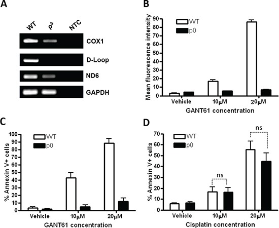
(A) Depletion of mitochondrial DNA in LO68 cells. Total DNA (20 ng) from wild-type (WT) and mitochondrial DNA-depleted ρ0 LO68 cells were subjected to PCR amplification using primers that were designed from specific regions of the mitochondrial DNA coding for COX1, D-loop and ND6. GAPDH was included as a control for nuclear DNA-encoded gene. NTC, no template control. (B) Detection of mitochondrial superoxide on GANT61 treatment. WT or ρ0 LO68 cells were treated with 10–20 μM GANT61 or vehicle for 48 h. The cells were then stained with mitoSOX red, and the fluorescence was measured by flow cytometry. Bar graph represents the increase in the mean fluorescence intensity of mitoSOX red-positive cells measured in each experimental condition. Values are the average of independent measurements (mean ± SEM; n = 3). (C) ρ0 LO68 cells are resistant to GANT61-induced apoptosis. WT or ρ0 LO68 cells were treated with 10–20 μM GANT61 for 48 h. Apoptosis was then measured by annexin-V/7AAD assay. Data represent the mean ± SEM of three independent experiments. (D) No apparent difference was observed in the sensitivity to apoptosis by cisplatin in WT or ρ0 LO68 cells. Cells were treated with 10–20 μM Cisplatin for 48 h. Apoptosis was then measured by annexin-V/7AAD assay. Data represent the mean ± SEM of three independent experiments. ns, not significantly different from untreated control cells.