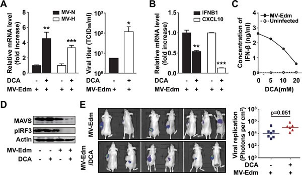
Figure 3. DCA promotes viral replication by disrupting MAVS-mediated anti-viral immune responses.

(A) U251 cells were infected with MV-Edm (MOI = 0.2) in the presence or absence of DCA (5 mM) for 24 h, then the total RNA in cells was harvested for the determination of viral genes encoding H and N proteins by qRT-PCR (left panel), or the supernatant was harvested for determination of TCID50 on Vero cells (right panel). Similar results were obtained in two independent experiments. (B) U251 cells were infected with MV-Edm (MOI = 0.2) in the presence or absence of DCA (5 mM) for 24 h, then the expression of IFNB1 and CXCL10 mRNA was determined by qRT-PCR. Means + SD of triplicates are shown. Similar results were obtained in three independent experiments. (C) U251 cells were infected with MV-Edm (MOI = 0.2) in the absence or presence of DCA (5, 10, or 20 mM) for 12 h, and supernatants were then harvested, and the protein level of IFN-β was measured by ELISA. (D) U251 cells were treated with DCA (5 mM), MV-Edm (MOI = 0.2), MV-Edm combined with DCA, or left untreated, and cultured for 24 h. Cell lysates were then harvested for immunoblotting against MAVS or pIRF3; β-actin was used as a loading control. A representative result from two independent experiments is shown. (E) U87 cells were inoculated subcutaneously into Balb/c nude mice. When tumors reached a palpable size, one group of mice received DCA (70 mg/L) in the drinking water for 10 d (n = 6). Another group was left untreated (n = 7). Then both groups of mice were injected with MV-Edm-Luc (4 × 10⁵ pfu per mouse) via tail vein. Luciferase activity was monitored by an in vivo luminescence imaging system 72 h after virus injection (left panel). Photons per cm2 tumor were quantified (right panel). * p < 0.05, ** p < 0.01, *** p < 0.001.
