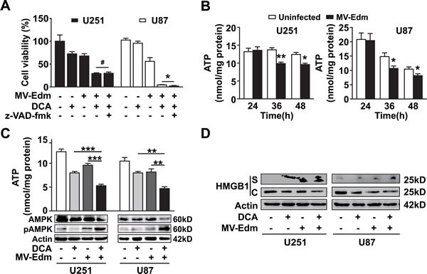
Figure 5. Necrosis contributes to MV-Edm/DCA mediated oncolysis by accelerated bioenergetics exhaustion.

(A) U251 and U87 cells were treated with DCA (5 mM), MV-Edm (MOI = 0.2), MV-Edm combined with DCA in the presence or absence of z-VAD-fmk (80 μM), or left untreated. Cell viability was determined by trypan blue exclusion 60 h post-treatment. Similar results were obtained in two independent experiments. (B) ATP content was determined in cell lysates harvested from U251 and U87 cells infected with MV-Edm at an MOI of 0.2 for 24, 36, or 48 h. Untreated cells were used as a negative control. Means + SD of triplicates are shown. Similar results were obtained in three independent experiments. (C) U251 and U87 cells were treated with DCA (5 mM), MV-Edm (MOI = 0.2), MV-Edm combined with DCA, or left untreated for 48 h. Cell lysates were then harvested for determination of ATP content (upper panel), or for immunoblotting against AMPK and phosphorylated AMPK (lower panel). Similar results were obtained in three independent experiments. (D) U251 and U87 cells were treated with MV-Edm (MOI = 0.2), DCA (5 mM), MV-Edm combined with DCA, or left untreated for 48 h. Cell lysates (C) and supernatants (S) were harvested for immunoblotting against HMGB1. β-actin was used as a loading control. Similar results were obtained in three independent experiments. * p < 0.05, ** p < 0.01, *** p < 0.001, # p > 0.05.
