
Figure 3. Casnosic acid induced down-regulation of c-FLIP and Bcl-2 expression at the post-translational levels.
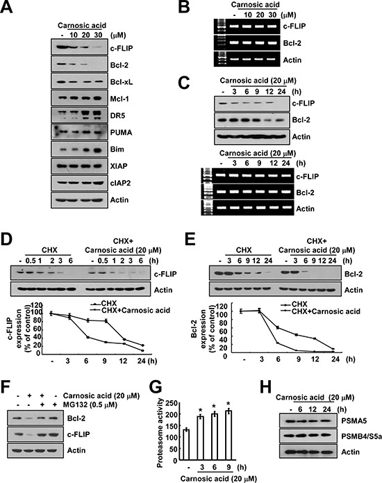
(A) Caki cells were treated with the indicated concentrations of carnosic acid for 24 h. The protein expression levels of c-FLIP, Bcl-2, Bcl-xL, Mcl-1, DR5, PUMA, Bim, XIAP, cIAP2 and actin were determined by Western blotting. The level of actin was used as a loading control. (B and C) Caki cells were treated with the indicated concentrations of carnosic acid for the indicated time periods. The mRNA and protein expression levels of c-FLIP and Bcl-2 were determined by RT-PCR (B and C) and Western blotting (C), respectively. The level of actin was used as a loading control. (D and E) Caki cells were treated with or without 20 μM carnosic acid in the presence of cyclohexamide (CHX) (20 μg/ml) for the indicated time periods. The protein expression levels of c-FLIP, Bcl-2, and/or actin were determined by Western blotting. The level of actin was used as a loading control. The band intensities of c-FLIP and Bcl-2 protein were measured using the public domain JAVA image-processing program ImageJ. (F) Caki cells were pretreated with 0.5 μM MG132, and then added 20 μM carnosic acid for 24 h. The protein expression levels of c-FLIP, Bcl-2 and actin were determined by Western blotting. The level of actin was used as a loading control. (G) Caki cells were treated with 20 μM carnosic acid for the indicated time periods. The cells were lysed, and proteasome activity was measured as described in the Materials and Methods section. (H) Caki cells were treated with 20 μM carnosic acid for the indicated time periods. The protein expression levels of PSMA5, PSMD4/S5a and actin were determined by Western blotting. The level of actin was used as a loading control. *p < 0.01 compared to the control.