
Figure 5. Carnosic acid induced ATF4 and CHOP-mediated DR5, Bim, and PUMA expression.
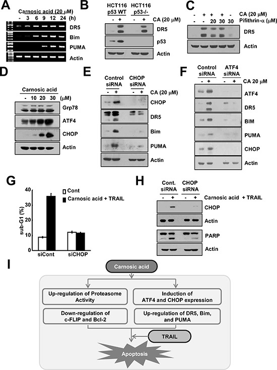
(A) Caki cells were treated with 20 μM carnosic acid for the indicated time periods. The mRNA expression levels of DR5, Bim, and PUMA were determined by RT-PCR. (B) p53 wild-type and p53−/− HCT116 cells were treated with 20 μM carnosic acid for 24 h. The protein expression of DR5, p53 and actin were determined by Western blotting. The level of actin was used as a loading control. (C) Caki cells were pretreated with the indicated concentrations of pifithrin-α, and then treated with 20 μM carnosic acid for 24 h. The protein expression of DR5 and actin were determined by Western blotting. The level of actin was used as a loading control. (D) Caki cells were treated with the indicated concentrations of carnosic acid for 12 h. The protein expression of Grp78, ATF4, CHOP, and actin were determined by Western blotting. The level of actin was used as a loading control. (E and F) Caki cells were transiently transfected control (Cont. siRNA), CHOP siRNA (E), or ATF4 siRNA (F). Twenty-four hours after transfection, cells were treated with 20 μM carnosic acid for 24 h. The protein expression levels of CHOP, ATF4, PARP, DR5, Bim, PUMA and/or actin were determined by Western blotting. The level of actin was used as a loading control. (G and H) Caki cells were transiently transfected control (Cont. siRNA) or CHOP siRNA. Twenty-four hours after transfection, cells were treated with 20 μM carnosic acid and 50 ng/ml TRAIL for 24 h. The level of apoptosis was analyzed by the sub-G1 fraction using flow cytometry (G). The protein expression levels of CHOP, PARP and actin were determined by Western blotting. The level of actin was used as a loading control (H). (I) Schematic models of carnosic-mediated TRAIL sensitization.