
Figure 6. MF-1 and DC101 suppressed metastasis of ESCC cells.
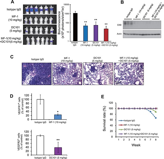
(A) Metastasis of ESCC cells was quantified by bioluminescence imaging at the 8th week after injection of KYSE150-Luc cells. (B) Western blot analysis of human-specific cytokeratin 8 protein in the lung extracts of 3 representative mice in each group. (C) Histological evaluation of lung metastasis (hematoxylin and eosin staining). (D) Flow cytometric analysis showed that MF-1 and DC101 significantly reduced the percentages of VEGFR1+ and VEGFR2+ cells, respectively, in the lungs. (E) The survival rates of mice over an 8-week period after introduction of cancer cells. Bars, SD; *, P < 0.05; **, P < 0.01, compared with isotype IgG-treated mice.