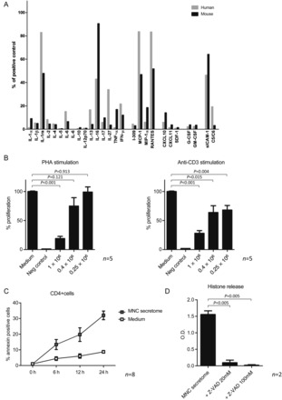
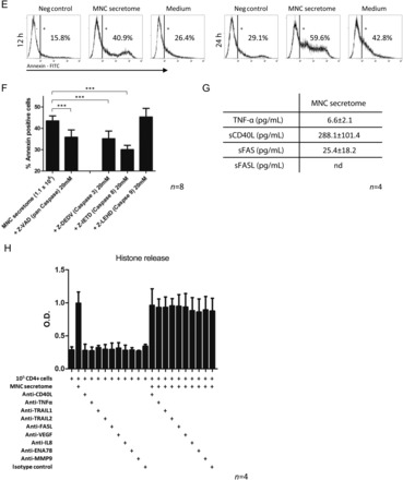
Figure 3.


Results of cytokine arrays of mononuclear cell secretome obtained from cultured mouse splenocytes and mononuclear cell secretome obtained from cultured human peripheral blood mononuclear cell are depicted in (A). Both preparations were comparable regarding their secreted products. (B) The proliferative response of purified human CD4+ cells in the presence or absence of mononuclear cell secretome. CD4+ cells stimulated with phytohaemagglutinin or anti-CD3 showed lower proliferation rates when treated with mononuclear cell secretome. Unstimulated, purified CD4+ cells or a commercially available T cell line (JURKAT) undergo apoptosis in the presence of mononuclear cell secretome as shown by flow cytometry (C and E) and by histone release assay (D). This effect was partially reversible by pre-incubation with a caspase-3 and a caspase-8 inhibitor, indicating an external pathway-mediated effect (F). Interestingly, known pro-apoptotic cytokines were only found in marginal concentrations in the mononuclear cell secretome (G). Blocking pathways associated with apoptosis by neutralizing antibodies had no effect on mononuclear cell secretome-induced histone release (H).
