. 2014 Aug 21;3:e03528. doi: 10.7554/eLife.03528

Figure 1. Poly-Ribo-Seq of small and large polysomes.

(A) Venn diagram categorising annotated Drosophila smORFs as corroborated or uncorroborated based on evidence (FlyBase) from two out of three of: GO molecular function term assignment (green), peptidomic evidence (blue), and conservation outside of insects (red). Based on this, out of the total of 829 annotated smORFs, 665 are uncorroborated, and 494 have no evidence of translation. (B) Schematic of Poly-Ribo-Seq with representative UV absorbance profile for sucrose density gradient. Small (purple) and large (blue) polysomes are separated and subject to ribosome footprinting. (C) Composite plot from all FlyBase protein-coding genes of Poly-Ribo-Seq read counts across mRNAs in the vicinity of start (upper) and stop codons (lower) in small polysomes. (D) Median translational efficiencies of CDS, 5′ and 3′-UTR regions for all protein-coding genes, error bars represent SE.

DOI: http://dx.doi.org/10.7554/eLife.03528.004

Figure 1.

Figure 1—figure supplement 1. Poly-Ribo-Seq of small and large polysomes.

Figure 1—figure supplement 1.

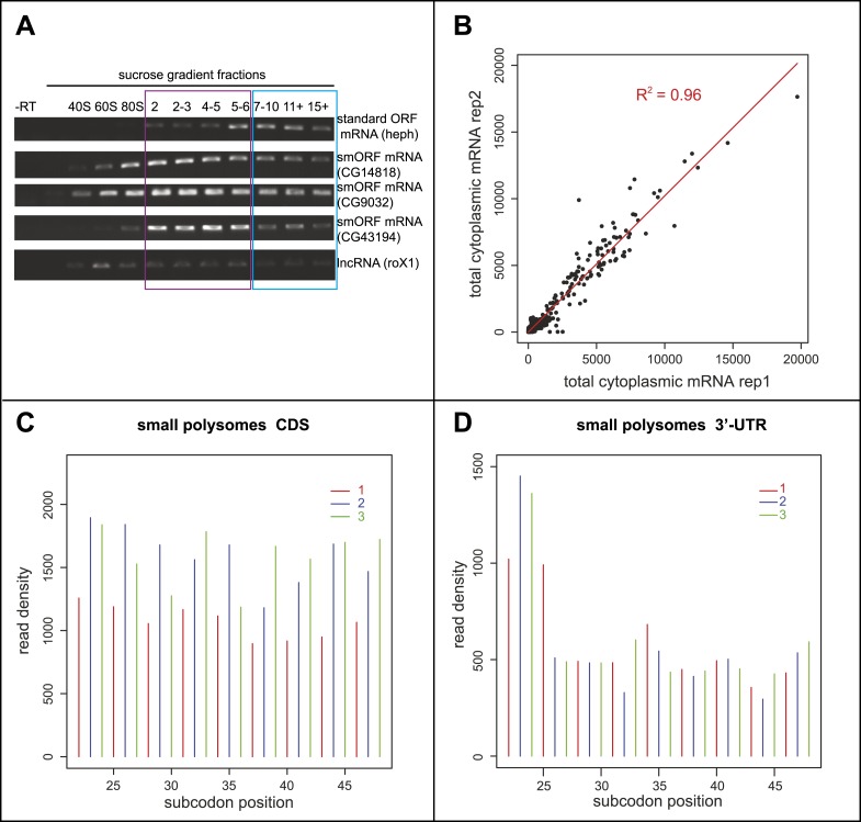
(A) RT-PCR of RNA recovered from sucrose gradient fractions for one standard ORF mRNA (heph), three annotated smORF mRNAs (CG14818, CG9032, and CG43194) and one long non-coding RNA (roX1), with -RT control. Fractions corresponding to small (purple, 2-6 ribosomes) and large (blue, 7 or more ribosomes) polysomes are indicated. (B) Read densities (RPKM) from two biological replicates of the total cytoplasmic mRNA control exhibit very high correlation (R2 = 0.96). (C and D) Read density plots showing phasing of ribosome footprinting reads in triplets corresponding to codons in CDS (C) and an absence of triplet phasing in 3′-UTRs (D) (small polysome data).

Figure 1—figure supplement 2. Schematic interpretation of Poly-Ribo-Seq.

Figure 1—figure supplement 2.

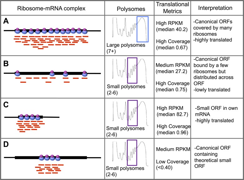
Schematic summary of characterised (AC) and theoretical (D) translation scenarios. Diagrams of ribosome–mRNA complexes are shown along with the polysome fraction in which it is detected, translational metrics and interpretation of this information, for (AC) long canonical ORFs, (C) smORFs, and (D) canonical ORF containing a theoretical small ORF.