. 2014 Sep 19;3:e03145. doi: 10.7554/eLife.03145

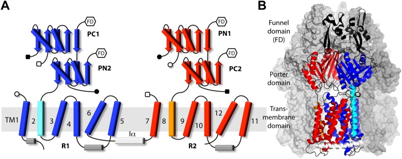
Figure 1. Topology and secondary structure representations of the AcrB protomers.

(A) Topology of the AcrB protomers in the transmembrane (TM) and porter domains. Our analysis indicates that the TM domain consists of two 5-helix parallel repeats, referred to as R1 and R2, and two flanking helices, TM2 and TM8. R1 (blue) is connected to R2 (red) through helix Iα (white), which lies parallel to the cytoplasmic face of the membrane. The porter domain also consists of two repeats of two subdomains each, namely PC1 and PN2 (blue), and PN1 and PC2 (red). TM2 (cyan) connects R1 to PN2, whereas TM8 (orange) links R2 to PC2. Other connections among porter sub-domains and R1/R2 are indicated by open/closed squares/circles. Hexagons indicate the connections between the porter domain and the funnel domain (FD), not drawn in this scheme. (B) Cartoon representation of the secondary structure of an AcrB protomer, in the context of the complete trimer. The other two protomers are drawn as a molecular surface (gray). Different sub-domains are colored as in (A), with inter-connecting loops and the FD shown in black.

DOI: http://dx.doi.org/10.7554/eLife.03145.003

Figure 1.

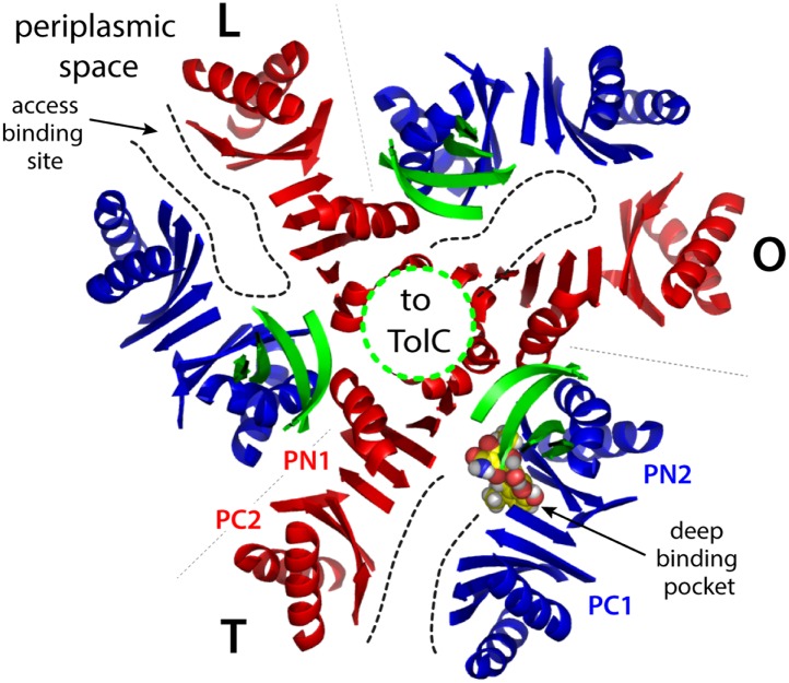
Figure 1—figure supplement 1. Cartoon representation of the porter domain of AcrB in the asymmetric state, viewed from the outer membrane, along the perpendicular to the inner membrane.

Figure 1—figure supplement 1.

Each of the protomers adopt a distinct conformation, namely L, T and O; gray dashed lines indicate the protomer interfaces. Subdomains PC1/PN2 and PN1/PC2 are colored in blue and red, respectively, as in Figure 1. A minocycline molecule is bound in the T state, in the deep binding pocket between PC1 and PN2. Substrates can also bind more superficially, in the L state. Black dashed lines indicate access pathways from the periplasmic space into the drug-binding sites, in the L and T states, and from the binding site to the central funnel (green dashed line) within the funnel domain, in the O state. A fragment of this funnel domain is shown with green cartoons; this funnel is open to the lumen of the TolC channel, which traverses the outer membrane of E. coli.

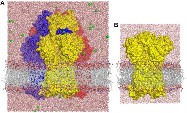
Figure 1—figure supplement 2. Molecular simulation systems employed in this study.

Figure 1—figure supplement 2.

(A) Asymmetric AcrB trimer (residues 1 to 1044 per protomer) with one minocycline bound to the T state, embedded in a POPC lipid membrane and solvated by water. The system is enclosed in a periodic box of dimensions ∼150 × 150 × 160 Å. The molecular system includes ∼370,000 atoms. AcrB is shown as a van-der-Waals surface, with the L, T and O states in blue, yellow and red, respectively. Lipid and water molecules are represented with lines and sticks; free Na+ ions, added to neutralize the total charge of the system, are shown as green spheres. (B) Free-energy simulations were carried out on a construct of the individual AcrB protomers, comprising the transmembrane and porter domains only, embedded in a POPC lipid membrane. The figure shows the T state; L and O states were prepared similarly. Each of these systems consist of ∼101,000 atoms, enclosed in a rectangular box of dimensions ∼90 × 90 × 120 Å.