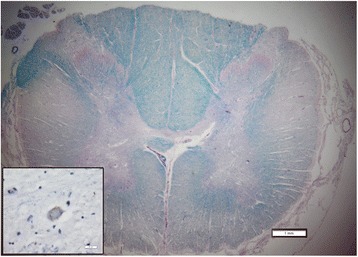
Figure 8.

Representative cross section of the spinal cord in the region of transplantation stained with luxol fast blue and periodic acid Schiff. There is no apparent disruption of tissue due to injection. Note the degeneration of the cortico-spinal tracts (“lateral sclerosis”). The inset demonstrates a phosphorylated TDP43 inclusion in a remaining motor neuron. Scale bars are 1 mm for the low power and 20 microns for the inset.
