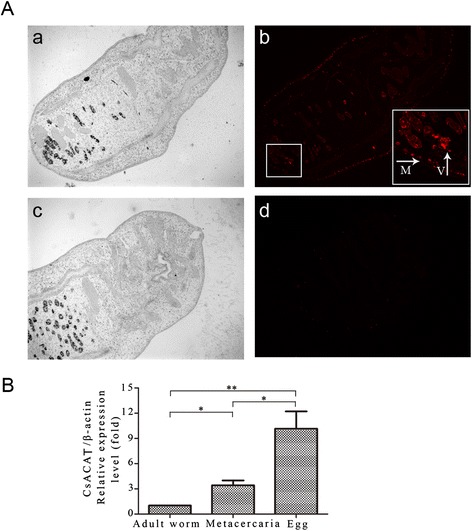
Figure 2.

Expression pattern of Cs ACAT. (A) Immunolocalization of CsACAT in C. sinensis adult. Panel a and b, sections treated with anti-rCsACAT serum and specific fluorescences distributed in the vitellarium and sub-tegumental muscular layer of the adult worm; Panel c and d, sections treated with naive serum. No fluorescence was detected. M, sub-tegumental muscular layer; V, vitellarium. Magnification for the adult worm was × 100. (B) mRNA level of CsACAT at different developmental stages of C. sinensis by quantitative real-time PCR. The specific mRNA fragment of CsACAT was observed among the stages. The quantities were normalized with Cs β-actin and analyzed by means of the 2−ΔΔCt ratio.
