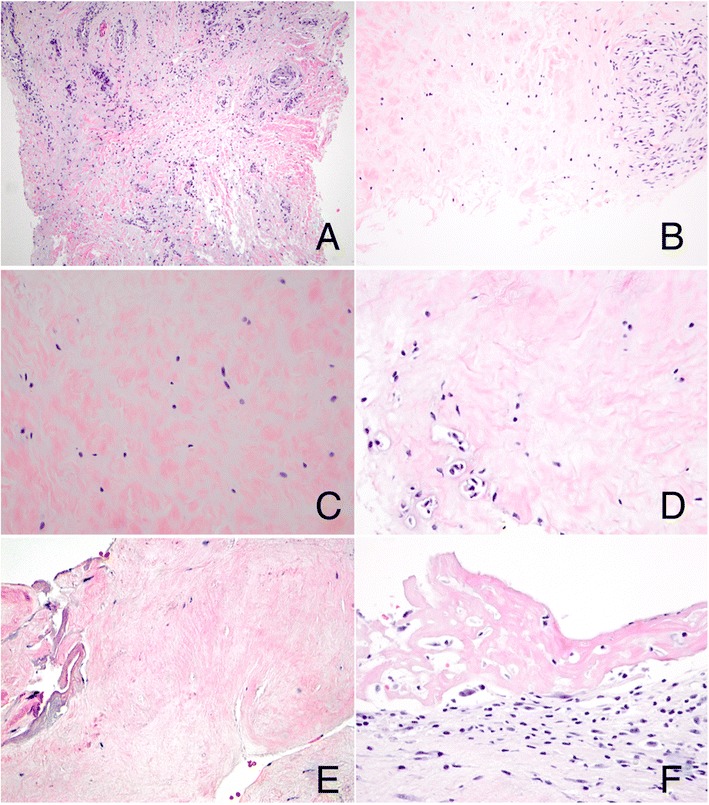
Figure 4.

Morphologic evaluation of dactylitis. Both the affected finger and toe showed tenosynovial tissue with prominent vascularity and excess myxoid extracellular matrix (A, finger, original magnification, 100×); additionally, fibrocartilage showed a prominent myxoid component (B, toe, original magnification, 200×; C, toe, original magnification, 400×), areas of binucleation (D, toe, original magnification, 400×), and focal hypocellularity and coagulative necrosis (E, finger, original magnification, 400×). Fibrinous synovitis was also seen (F, finger, original magnification, 400×).
