Figure 2.

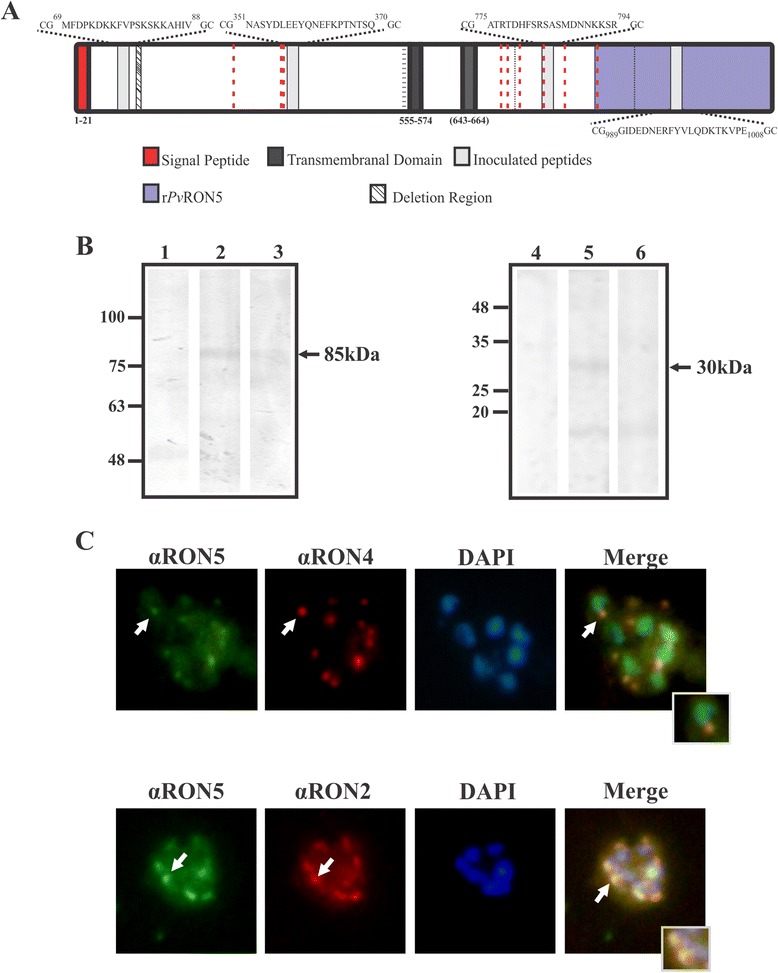
Pv RON5 expression in Plasmodium vivax schizonts. (A) Representation to scale of the P. vivax RON5 protein. The signal peptide and the two transmembrane domains predicted by bioinformatics tools and the localization and sequence of the linear B-cell epitope peptides selected for polyclonal antibody production are shown. Comparative analysis of amino acid sequences from different P. vivax strains revealed the deletion of a nine-residue-long region; black dotted lines show synonymous and non-synonymous changes. Aligning P. vivax, P. falciparum and P. knowlesi RON5 revealed nine conserved cysteines (red dotted lines). The recombinant protein (rPvRON5) produced in E. coli is shown in purple. (B) PvRON5 expression in VCG-1 strain schizont lysate. Lane 1, pre-immune serum 60; lane 2, immune serum 60; lane 3, immune serum 60 pre-incubated with peptides 36930 and 36927; lane 4, pre-immune serum 2; lane 5, immune serum 2 and lane 6, immune serum 2 pre-incubated with peptide 39274. (C) PvRON5 sub-cellular localization in P. vivax-infected RBCs in schizont stage. Green shows serum reactivity for PvRON5, having a dotted pattern similar to that observed for PvRON4 and PvRON2 (red). The arrows show the dotted pattern and the overlaying of the images (merging). DAPI (4′,6-diamidino-2-phenylindole) was used for staining the parasite nucleus.
