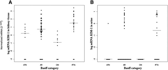
Figure 1.

The dot-plot representation graphics showing the medians and distribution of the quantification levels of normalized KIM-1 mRNA (2 -ΔΔCT ) according to the Banff diagnostic groups. Panel A: kidney tissue. Panel B: urinary sediment cells. ATN = acute tubular necrosis; AR = acute rejection; CIN = calcineurin inhibitor-induced nephrotoxicity; IFTA = interstitial fibrosis and tubular atrophy.
