
Figure 1.
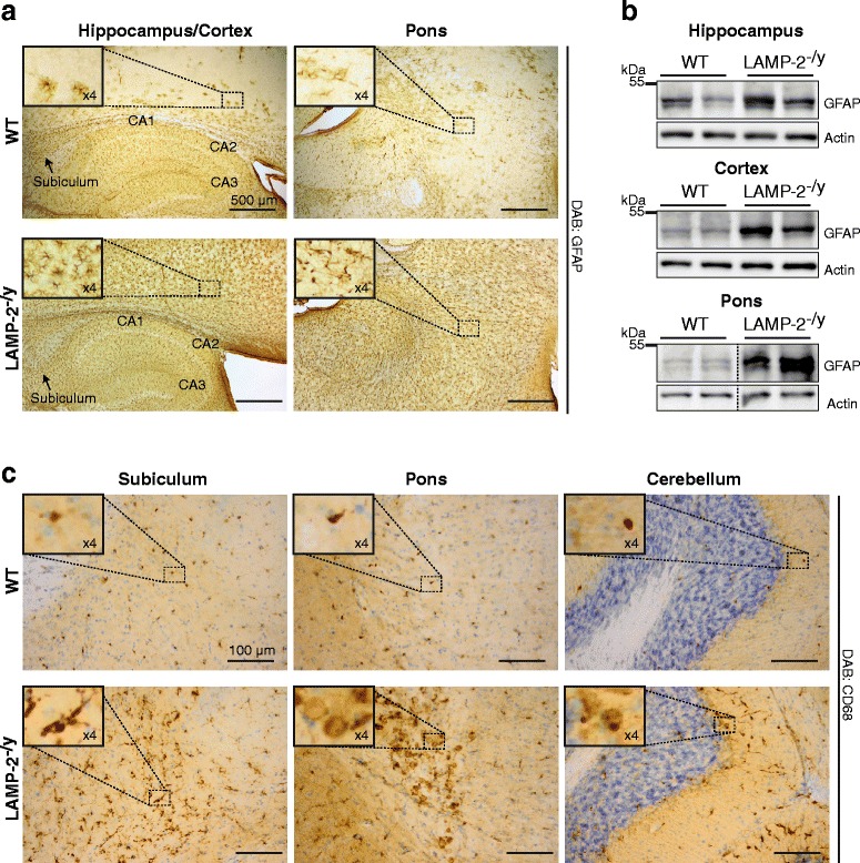
Neuropathological changes in the absence of LAMP-2 expression. Representative histological brain sections from LAMP-2-deficient (LAMP-2-/y) mice and their wild-type (WT) littermates (zoomed images shown in insets). (a) Astrogliosis in LAMP-2-/y brain visualized with the aid of GFAP immunological staining. (b) Immunoblotting of brain lysates showing GFAP expression levels in WT and in LAMP-2-/y brain tissue. (c) Microgliosis observed in LAMP-2-/y animals via CD68 immunological staining (sections were costained with Nissl).